Showing posts with label digital dental radiograph machine. Show all posts
Tuesday, August 21, 2012
Dental surprise!
One of my patients, Tazo, was having a hard time chewing her food so she was brought in for an exam. Her 108 (really big upper molar had some gum recession and purulent (abcess material) along the gum line. We scheduled her for surgery.
Well, today was the day. We took a radiograph to see what was going on with the bone around the tooth and to see if there were any tooth root abcesses. I was not expecting to see this picture. The entire back root was gone, as was the bone from infection. It also usually takes anywhere from 20-40 minutes to remove this tooth but it only took half the time. The biggest problem was getting enough gum tissue to cover the sockets once I got all the "debris" out of them.
Here is the after picture with the area sutured close. No more wiggly, painful tooth! Plus, her breath doesn't clear a room out now.
Friday, December 9, 2011
Surprise!

I have learned to expect the unexpected when doing dentals on my patients. The past few dentals I have done, have proven that beyond a reasonable doubt.
Duffy was one of my dental surgery patients. When I saw the small area of gingivitis on the premolar tooth (307), I took a dental radiograph. I was surprised at the extensive loss of bone around the roots of the teeth. I was expecting a little loss around the one tooth, not all three teeth. The two yellow lines represent where the bone is supposed to be holding the teeth in place.
Before I had dental rads, I would likely have only known to remove that one tooth. The red lines show the fused roots of 304 and 305. (Luckily I watched Sesame Street as a kid and learned how to count. All the teeth have assigned numbers.) Knowing that important piece of information allowed me to adjust how I drilled to expose the these roots and remove them easily.
Due to the extent of Duffy's dental disease, he went home with about eight teeth. Now don't worry. If our pet's can eat with that kind of dental disease, just think how well they do when they have healthy gums!
Wednesday, February 16, 2011
You ate what???

When I went to work this morning, I only had one surgery scheduled. That meant I would have time to do all the paperwork I’ve been putting off. I almost got myself two coffees at Tim’s to make sure I could stay awake. Turns out I wouldn’t need the one I had!
While PJ and Kelly (my AHTs) were prepping my neuter for surgery, I had a walk-in appointment. Juno had not been feeling well for a few days and was progressively getting worse. She might have eaten a hockey sock or two as she had vomited one up earlier. Entering the room showed a very different dog than I was used to handling. My happy monkey was now very depressed with a painful abdomen.
We did radiographs and in addition to the odd intestinal pattern there was also loss of detail in the cranial abdomen. The decision was to immediately go to surgery. I was so proud of my staff getting me in so quickly and efficiently. Kelly was monitoring and PJ scrubbed in to help.
The foreign body extended from the pylorus (stomach) through to the jejunum. As you can see from the picture, the intestines I’m holding are abnormal. They should look like the ones on the left of the picture. The problem area has the intestines plicated (squished together tightly). This caused a 12 inch section to die from lack of blood supply.
The foreign body was about 24 inches long. I had to wait until the surgery was done and Juno recovered before I could rinse the “thing”. It turned out to be a part of a dishcloth.
Sunday, January 2, 2011
No Bones Please!

On a fairly regular basis, I have owners tell me that one of their dog’s favorite chew toys is a bone. Unfortunately I have seen all types of bones cause problems. At this point in my career, I don’t care if it’s the biggest knuckle bone that’s been boiled. It can still cause issues.
The most common issue is gastrointestinal (GI). Bones can get caught in the esophagus, trachea, stomach and/or intestines. If the bone(s) poke through the stomach or intestines it can cause peritonitis. Peritonitis is a very difficult to treat bacterial infection of the abdomen. Bones will often scrape the lining of the intestines causing bleeding. Vomiting and diarrhea are also seen and may indicate surgery is needed.
Early in my career, I literally saved a patient by doing a rectal exam. A couple brought in a very lethargic 85lb dog. On physical exam, his abdomen was painful but all he could really do was groan. His history included getting a new bone every weekend. They swore he was never able to get any pieces off the bone. Well, the rectal found a piece of bone wedged sideways in the pelvis. I was able to gently turn and remove it. A follow up call found him back to his usual self.
The most recent bone case involved a goofy Labrador retriever (as an owner as well, what else can you say about them?). She had been seen a couple days before at the emergency clinic for vomiting and diarrhea caused from eating a bone. Antibiotics and stomach protectants were dispensed. When I saw her, she hadn’t improved very much so we took the radiograph. At least bones show up easily! The shards of bone were in the large intestine so we knew it was just a short matter of time before they would pass and she could continue to heal. She was one of the lucky ones.
Sunday, December 19, 2010
Surgical Surprises

This week we had a couple of surprises when it came to our surgeries. The first one had the owners, staff and I laughing. A new client brought in the families new 16 week old kitten to be neutered. They had been told it was a male but the kids wanted to call him Dolly. When we are admitting for surgery we always double check just to be sure. Well…..turns out he was actually a she and the kids had found the perfect name!
The other neat find was when we were getting ready to do a spay on a 6 month old small breed dog. My AHT, Kelly, asked me to take a quick peak at the mouth as there were some retained baby teeth. There were five upper baby incisors that needed to come out but the bottom teeth were the most intriguing. There were only three adult incisors (supposed to have six) and the baby canine teeth hadn’t fallen out which was causing the adult teeth to erupt to the inside of the mouth.
This can become a major problem. If we did not remove the baby teeth, the adult teeth would continue to erupt improperly and would end up damaging the hard palate. My mouth hurt just thinking about what could happen. I took a quick dental radiograph to make sure nothing else was going on in the lower jaw.
Once I was done with the routine spay, I extracted the baby teeth. This is the one time we do not suture the incisions closed. We want to give the adult teeth every chance to move outward to their normal position.
Sunday, November 7, 2010
Holy Ouch!!!
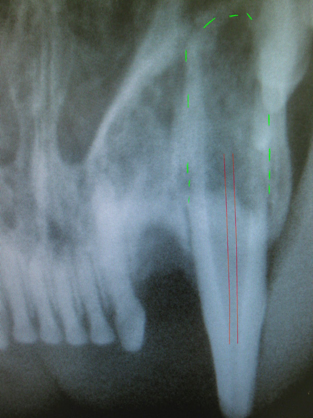
I had one of the neatest dental surgeries that I’ve had in ages. I’ve known Morris, a 10 year old DSH cat for the past few years. He has never been a “white smock” fan and since I tend to wear them…….. On his last physical exam, I noticed that he had fractured one of his upper canine teeth. The pulp was black so we needed to go forward with dental surgery.
Before removing teeth, I almost always take a radiograph. I want to see what shape the roots and bones are in. I was more than a little confused when I saw the radiograph for this tooth. Normally, the pulp of a mature cat or dog becomes a very narrow. The pulp on this one got so large the root basically disappeared.
I carefully elevated and drilled around the tooth root. I tried to be even more delicate than usual removing the root. I was astounded as the tooth was completely full of abscessed material. Then I had to use a special tool to scrape the rest of the abscess out of the pocket. The gums were sutured closed and my AHT, Kelly, finished the charting, cleaning and polishing.
I know that Morris will feel much better having that abscessed tooth out of his mouth. I wish that he would associate that feeling with the white smock.
Picture: the red line represents where the pulp should be and the green lines represent where the tooth root should be.
Wednesday, October 20, 2010
Joyous Miracles

Last week, one of my clients came in to have a radiograph done on her pregnant dog, Joy. This breeder is very responsible, she makes sure to have all of the appropriate tests done prior to breeding (OFA certification of hips, Optigen test for PRA, eye certification and Baer testing for hearing). Her dogs have their championships and has litters maybe once per year or every other year. We were very happy to see four cute little puppies.
Right before surgery this morning, I received a call saying that Joy had started whelping (having pups) at 4:30 am. Her owner found that she was having weak, inconsistent uterine contractions. The second puppy arrived just after 6:30am. Since it was now almost 9am, I told her to bring Joy right in. Surgery was going to have to wait.
While waiting for her to arrive, I prepared the staff. We needed to have the exam room set up with blankets, small towels (for drying the puppies off), suture (tying off the umbilical cord and betadine to clean the end of the cord. The surgery room was also set up in case we needed to do an emergency caesarian section.
Upon arrival, both the owner and Joy appeared very tired. The two newborns looked strong. I did an internal vaginal exam but only got a mild Ferguson’s reflex (when the uterus is palpated, it should contract). Oxytocin was given to stimulate contractions and the last two finally arrived. Sighs of relief were heard all around when the squeals started up and the best part was watching the babies starting to suckle.
Thursday, August 5, 2010
Eva's input in dental surgery


When I invest in certain “toys” for my hospital, I tend to name them. Eva is my digital dental radiograph machine. I can have a radiograph (x-ray) within seconds and determine whether the tooth (or teeth) can be salvaged or need to be removed.
Many people assume that animals will always show pain and stop eating when they have a sore mouth. If only that were so. I am always amazed how “normal” they act even when there are slab fractures of the teeth, loose teeth or even abscesses of the tooth root. What gives me satisfaction are the phone calls from owners who are amazed because their dog or cat is acting happier once the dental surgery has been performed.
Bud is one of my more recent cases. Over the years, he has had a few dental procedures. During his biannual senior exam, I found that he had progressed to stage 2 out of 4 dental disease. This means he has moderate tartar and gingivitis. His family and I decided it would be a good idea to schedule bloodwork, then a thorough cleaning and polishing.
It turned out to be an excellent day to have Eva around. While one of my AHTs was charting and probing his teeth, she found a pocket around one of the teeth of the lower jaw. I decided to take a radiograph. It turned out that two teeth needed to be extracted. There was both bone loss and abscesses at the tooth roots. Without Eva, I would not have known how extensive the dental disease actually was in the mouth.
According to his owners, he was back to better than his normal self in just a few days. Bud also wanted to play fetch again, which they hadn't realized he'd stopped playing a number of months ago.
Subscribe to:
Posts (Atom)